What AI-Assisted Imaging Really Means for the Emergency Department

A working ER physician on the promise, the friction, and the quiet ways algorithms are already reshaping the triage bay.

By Dr. Marcus Halden, ER Physician & Contributor
7 min read
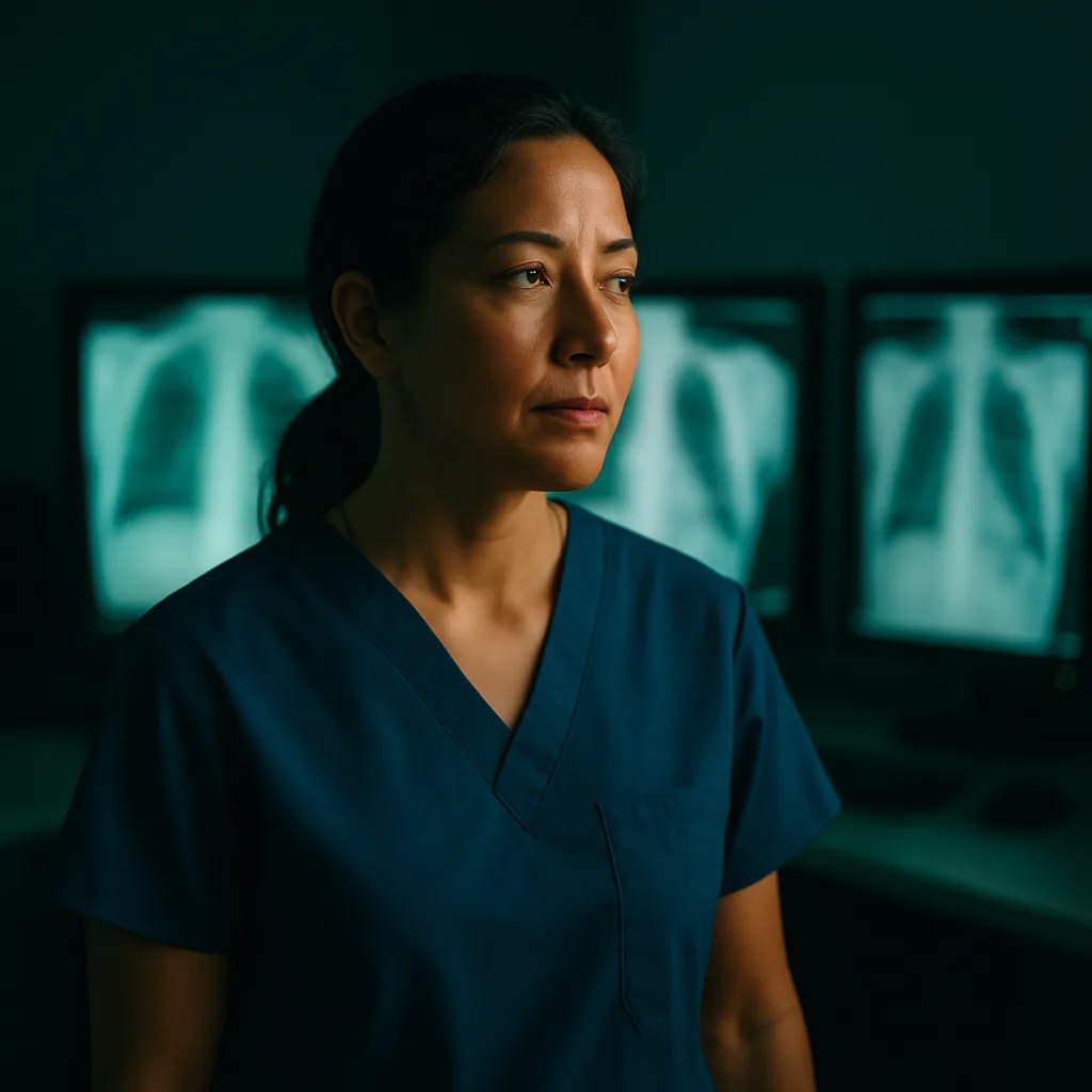
ER physician reviewing imaging at night

Field Notes / Vol. 07

A Letter From The Attending

The algorithm saw it first. I was still reading the chart.

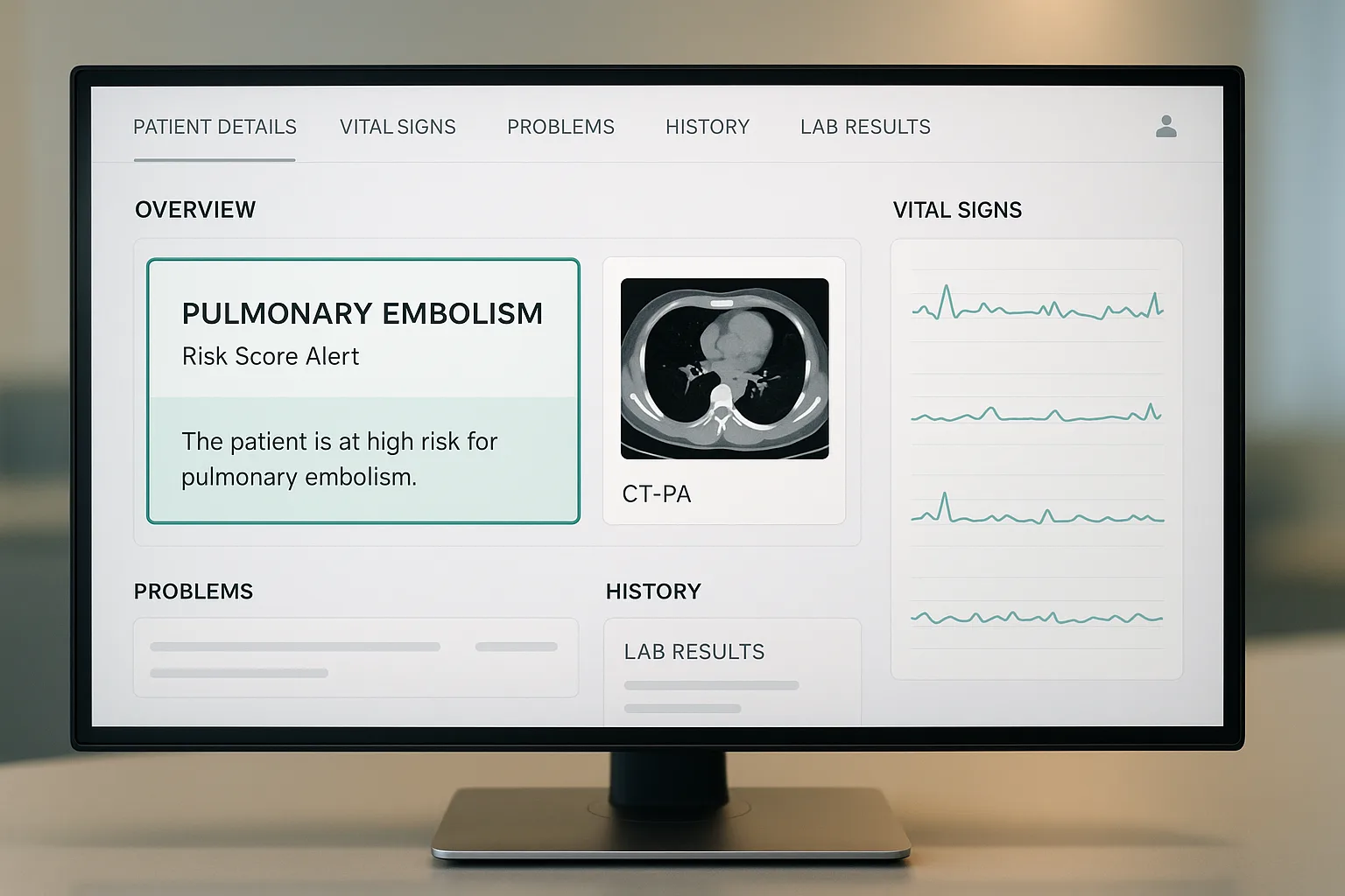
It was a Tuesday. 11:47 p.m. A 58-year-old woman, shortness of breath, vitals borderline unremarkable. Before I had finished her history, a soft chime on the workstation flagged a high-probability pulmonary embolism from the CT reconstruction. My resident looked at me. I looked at the screen. And something quietly shifted in the room.

I have spent the better part of two decades in emergency departments, and I will confess I came to artificial intelligence the way most of my colleagues did: skeptical, tired, and a little tired of being sold to. Every conference season, a new vendor promises the moon. Ambient scribes. Predictive sepsis. Autonomous triage. The demo runs beautifully in a Marriott ballroom. Then it hits the floor at 3 a.m. on a Saturday, and the promises begin to fray.

What actually ships to the department is rarely what the keynote described. The models are narrower. The integrations are clumsier. The alerts, God help us, are louder. In my first year with a decision-support tool bolted onto our EHR, I clicked through so many low-yield pop-ups that I stopped reading them. That is the honest arithmetic of alert fatigue, and no glossy case study will make it prettier.

Then there is the harder conversation, the one we have in the physician lounge and rarely in print: the trust gap. When a black-box output disagrees with my read, who owns the miss? What do I tell the family? What does the medical board tell me? These are not rhetorical questions. They are the reason many of my colleagues, good clinicians, careful thinkers, keep the new tools at arm's length.

AI is not replacing the physician. It is reshaping the cognitive load in ways the field is still learning to measure, and still learning to name.

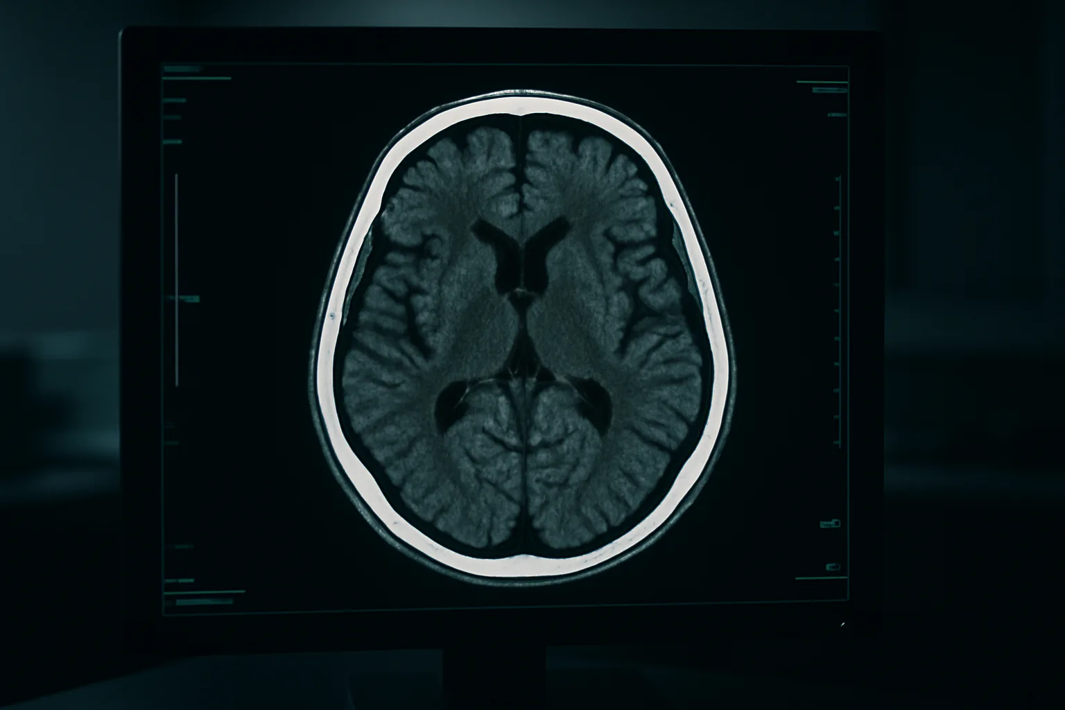
And yet. I have watched a chest X-ray triage model pull a subtle pneumothorax to the top of the worklist and shave forty minutes off a chest tube. I have watched a hemorrhage detection tool catch a small subdural on an overnight read when our on-call radiologist was three studies deep. I have watched time-to-CT-read in off-hours coverage drop in ways that, quietly, save lives without ever making a press release.

These are not moonshots. They are small, unglamorous wins, the kind that accumulate into better medicine when the technology is deployed with humility and the workflow is designed by people who have actually stood in a triage bay at shift change. That last part matters more than any benchmark.

I do not think AI will replace what I do. I do think it is quietly rewriting the cognitive geometry of the job: what I hold in working memory, what I delegate to the machine, what I still must own with my full attention. We are only beginning to build the vocabulary for it. Ameri-Med exists, in part, so that vocabulary is written by clinicians and patients, not just by the vendors selling us the tools.

I wrote this letter for the physicians, nurses, students, and patients who read this platform, because I think we should talk more honestly about what is happening at the bedside. Not the hype. Not the fear. The quieter, stranger truth in between.

Elena R. Marchetti, MD

Attending Physician, Department of Emergency Medicine
Associate Professor of Clinical Medicine
Contributing Editor, Ameri-Med
Where algorithms are already working

Three imaging workflows, live in emergency departments today. Not pilots. Not white papers. Real clinicians, real reads, real minutes shaved off the door-to-diagnosis clock.

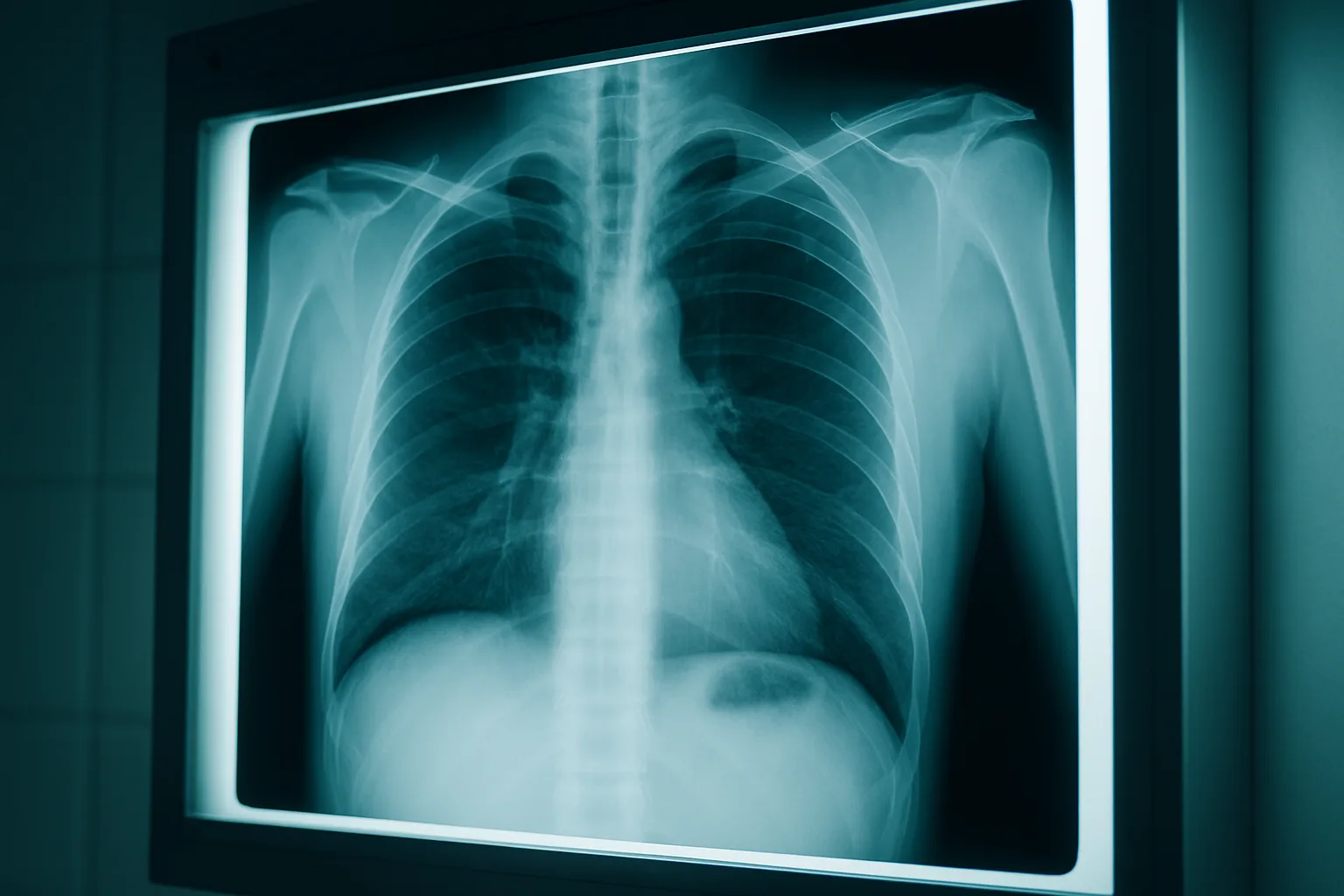
Chest X-ray on hospital lightbox
01 Deployed

Chest X-Ray Triage Support

AI flags potential pneumothorax and consolidation patterns in under 60 seconds, allowing nurses to escalate before physician review. What used to sit in a queue now surfaces the moment the plate lands.

02 Deployed

Intracranial Hemorrhage Detection

Deep-learning models trained on millions of CT slices surface suspected bleeds at the top of the read queue, compressing door-to-diagnosis time when every minute of ischemia matters.

CT brain scan on radiology workstation
EMR dashboard with pulmonary embolism risk alert
03 Deployed

Pulmonary Embolism Risk Stratification

Automated analysis of CT-PA scans cross-references prior imaging and vitals to produce a risk score surfaced directly in the EMR, giving the treating physician context before they've opened the study.

Abstract blurred hospital corridor at night with soft teal-toned ambient lighting, out-of-focus fluorescent glow, cinema

Dispatches from the floor

The algorithm doesn't make the call, I do. But it has quietly taken some of the loneliest decisions in medicine and made them feel a little less alone.
ER Attending Physician
Ameri-Med contributor

One of many voices from the physicians, nurses, and patients shaping the conversation around emerging medical technology.

Read more voices

FAQ · Clinical & Operational

Common Questions from the Floor

Straightforward answers to what emergency clinicians, radiologists, and administrators are actually asking about AI-assisted imaging in the ED.

Is AI imaging assistance FDA-cleared for emergency use?

Several platforms have 510(k) clearance for specific indications, including intracranial hemorrhage, large-vessel occlusion, and pulmonary embolism triage. Clearance scope varies by modality and vendor, so procurement teams should verify indication-by-indication rather than assuming blanket approval.

Who is liable when an AI-flagged finding is missed?

Liability remains with the ordering and interpreting physician. AI output is advisory, not diagnostic, and the medical record should reflect that the clinician made the final call. Institutional policies increasingly formalize this boundary in imaging protocols.

Does AI imaging slow down existing ED workflows?

Integration quality is the deciding factor. Poorly implemented tools generate alert fatigue and click burden that erode adoption within weeks. Purpose-built ED integrations, on the other hand, can measurably reduce time-to-read for time-critical findings by surfacing them directly in the existing PACS worklist.

How do smaller community EDs without 24/7 radiology coverage benefit most?

Off-hours triage support and priority queuing deliver the highest measurable impact in low-staffed settings. When a stroke or hemorrhage flag reaches the on-call clinician before the teleradiology read returns, minutes translate directly to preserved brain tissue and better outcomes.

Will radiologists and ER physicians lose roles to AI?

Evidence points to augmentation, not replacement. AI handles volume and pattern recognition at scale; clinicians handle judgment, clinical context, and the patient in front of them. The clinician-AI team consistently outperforms either alone in published ED studies.

Have a question we haven't covered? We publish physician-submitted responses weekly.

Submit a Question
The Dispatch

Stay Ahead of the Algorithms

Ameri-Med publishes physician-authored analysis on AI, imaging, and the tools reshaping emergency medicine. No vendor noise, just real clinical perspective.

No spam. Unsubscribe anytime. America's voice in emerging medical technology.